Protocol for NucleoLIVE™ Non-Toxic Dye
This is intended as a guide only.
In Brief Download the PDF of this protocol
NucleoLIVE™ Non-Toxic Dye is a ready-to-use, wash-free, fluorescent nuclear stain for live cell imaging. It is highly suited for long term 2D and 3D cell organoids imaging, and for monitoring precise tracking of cell proliferation, differentiation, and responses to stimuli over time. Demonstrates excellent cell viability, high signal-to-noise ratios, and robust segmentation in 2D and 3D models.
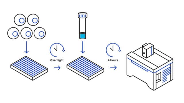
1. Protocol Overview

2. Content and Storage
| Product | Content | Storage | Stability |
|---|---|---|---|
|
NucleoLIVE™ Non-Toxic Dye
|
Diluted in 50 µl of DMSO
|
-20º C Delivered at room temperature Protect from light |
1 year
|
Table 1. NucleoLIVE™ Non-Toxic Dye Product Information
Intended Use: For research use only. Not for use in diagnostics or therapeutic procedures.
3. General Guidelines
Dye dilution and preparation
- Warm up the NucleoLIVE™ Non-Toxic Dye tube to room temperature before use to avoid condensation to form and water to get into the anhydrous dye solution
- Gently spin the tube before use to collect any dye solution that may remain near the cap
Quick Protocol
- Days prior: grow cells to the desired density on your preferred imaging-compatible substrate (coverslips, clear-bottom multi-well plates, etc.)
- Staining solution: Dilute the provided 1,000x nuclear stain in your usual culture medium
- Replace the cell medium with the prepared staining solution
- Incubate at 37°C, for 4h
- Live cells can be imaged without needing to wash out the excess probe in the media
Notes
- Probe concentration and incubation times are given as general guidelines and have been validated on MCF-7 breast cancer cells.
(1): Higher concentrations (1:500 dilution) can be used for harder-to-stain cell lines, or shorter labeling periods. Lower concentrations (1:2,000-5,000 dilutions) can be used when imaging live cells for longer periods of time (>24h).
(2): Incubation times with the probe should be validated for each cell line, especially when using higher dilutions.

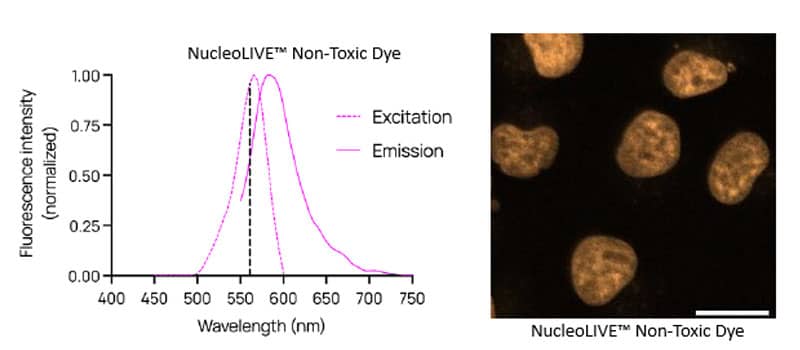
Figure 2. Excitation and Emission spectra of NucleoLIVE™ Non-Toxic Dye