Protocol for MortaLIVE™ Non-Toxic Dye
This is intended as a guide only; for full experimental details please read the reference provided.
In Brief Download the PDF of this protocol
MortaLIVE™ Non-Toxic Dye is a ready-to-use fluorescent dye to quantify live and dead cells and assess cytotoxicity in real-time. MortaLIVE™ Non-Toxic Dye is extremely simple to use, being added directly to the cell culture medium, and being imaged on standard imaging setups at multiple timepoints throughout the assay, allowing for real-time cytotoxicity monitoring over multiple days.
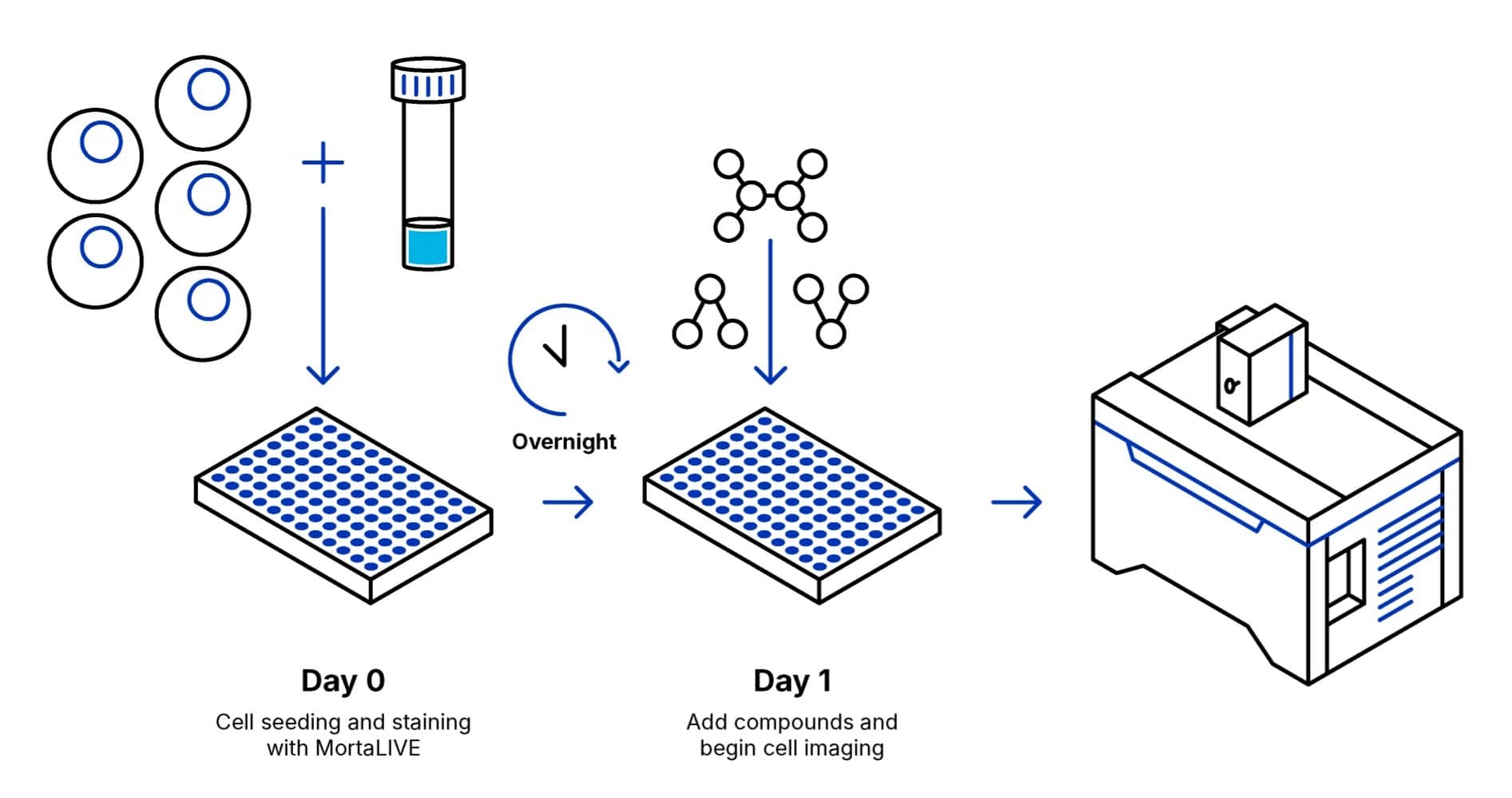
1. Protocol Overview

2. Content and Storage
| Product | Content | Storage | Stability |
|---|---|---|---|
|
MortaLIVE™ Non-Toxic Dye
|
Diluted in 50 µl of DMSO Recommended dilution is 1:1,000
|
-20º C Delivered at room temperature Protect from light |
1 year
|
Table 1. MortaLIVE™ Product Information
Intended Use: For research use only. Not for use in diagnostics or therapeutic procedures.
3. General Guidelines
MortaLIVE™ Non-Toxic Dye dilution and preparation
- Warm up the MortaLIVE™ Non-Toxic Dyes tube to room temperature before use to avoid condensation to form and water to get into the anhydrous dye solution.
- Gently spin the tube before use to collect any dye solution that may remain near the cap.
Cell culture protocol and cell viability testing with MortaLIVE™ Non-Toxic Dye
- Seed cells at desired density (typically to achieve 70-80% confluence) on imaging support, in cell culture medium.
- Add control compounds, test compounds and negative controls for phenotypes of interest.
Staining
- Dilute the provided 1,000x MortaLIVE™ Non-Toxic Dye in your usual culture Medium (e.g.: dilute 10 µL MortaLIVE™ Non-Toxic Dyes 1,000x in 10 mL culture medium).
- Replace the cell medium with the prepared staining solution.
- Incubate at 37°C, for at least 30 min.
Imaging
- Live cells can be imaged without needing to wash out the excess probe in the media.
NOTE:
Probe concentration and incubation times are given as general guidelines and have been validated on MCF-7 breast cancer cells.
MortaLIVE™ Non-Toxic Dye solution can also be prepared more concentrated, and added on top of the existing medium in the well (e.g.: prepare a 10x staining solution by diluting 1 μL MortaLIVE™ Non-Toxic Dye 1,000x in 100 μL medium, then add 11 μL 10x staining.)

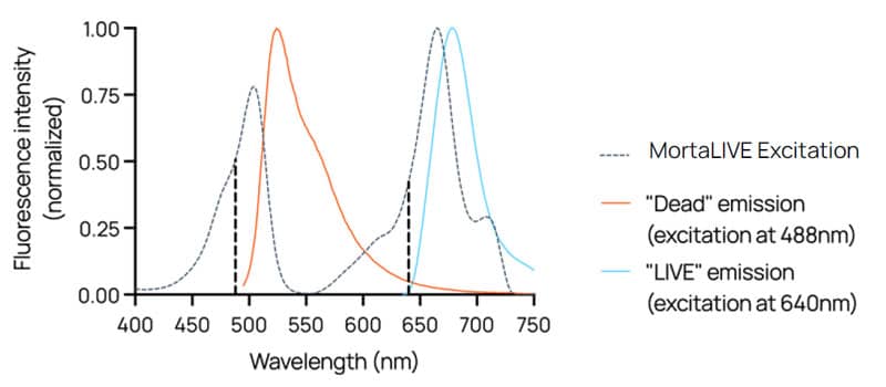
Figure 2. MortaLIVE™ Non-Toxic Dye spectrum
4. Image Examples
Quantifying Cytotoxicity Over Time in Kinetic experiments

Figure 3. MCF-7 cells stained with non-toxic MortaLIVE™ Non-Toxic Dye and with NucleoLIVE™ Non-Toxic Dye (live in red and dead in green channel, with dead also appearing aqua, and nuclei in blue). Cells are treated with increasing doses (left to right) of staurosporine (top row) and of thapsigargin (bottom row), and imaged at 48h.

Figure 4. Concentration-response curves at 48h using intensity data from the live and dead channels.