Protocol for ChromaLIVE™ Non-Toxic Dye
This is intended as a guide only.
In Brief Download the PDF of this protocol
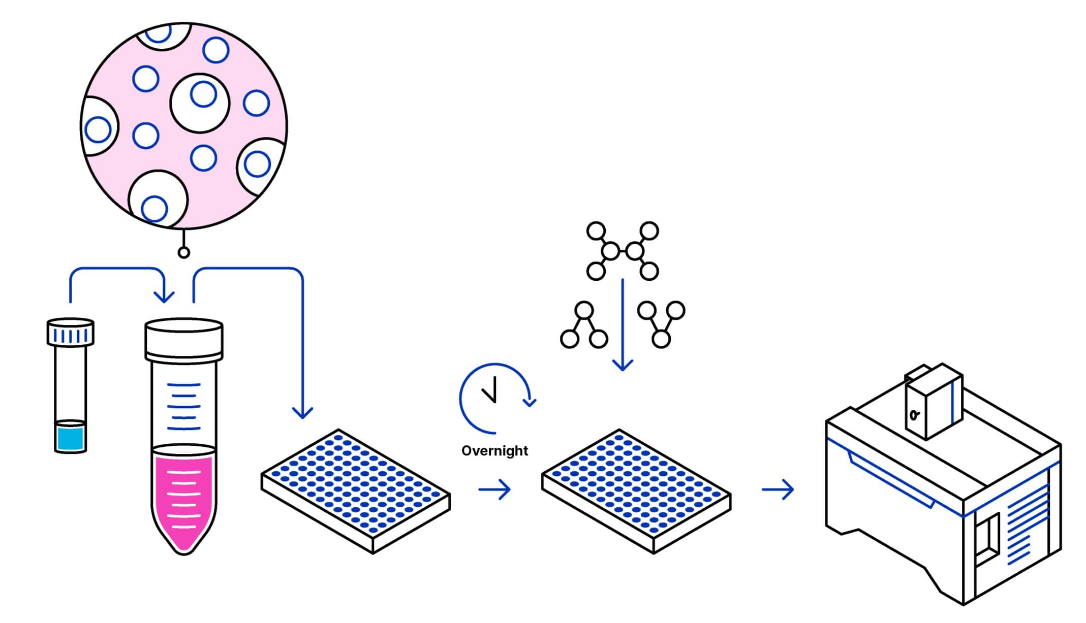
ChromaLIVE™ Non-Toxic Dye is a ready-to-use fluorescent probe for long term high content phenotypic screening of live cells. It allows live cell phenotypic imaging with a single dye enabling multi-organelle phenotypic readout to accurately differentiate cellular phenotypes stress and cell death pathways.
1. Protocol Overview

2. Content and Storage
| Product | Content | Storage | Stability |
|---|---|---|---|
|
ChromaLIVE™ Non-Toxic Dye
|
Diluted in 10 µl of DMSO
|
-20º C Delivered at room temperature Protect from light |
1 year
|
Table 1. ChromaLIVE™ Non-Toxic Dye Product Information
Intended Use: For research use only. Not for use in diagnostics or therapeutic procedures.
3. General Guidelines
ChromaLIVE™ Non-Toxic Dye dilution and preparation
- Warm up the ChromaLIVE™ Non-Toxic Dye tube to room temperature before use to avoid condensation to form and water to get into the anhydrous dye solution
- Gently spin the tube before use to collect any dye solution that may remain near the cap
- Dilute ChromaLIVE™ Non-Toxic Dye in preferred culture medium (1:1000 the provided solution)
- Vortex thoroughly
Cell culture protocol and compound testing, with ChromaLIVE™ Non-Toxic Dye
- Seed cells at desired density (typically to achieve 70-80% confluence) on imaging support, in previously prepared culture medium with ChromaLIVE™ Non-Toxic Dye
- Add control compounds, test compounds and negative controls for phenotypes of interest
NOTE: ChromaLIVE™ Non-Toxic Dye can also be added after cell seeding and compound addition. In that case, we recommend running a preliminary imaging test to validate the staining kinetics for your cells. As reference, ChromaLIVE™ Non-Toxic Dye staining stabilizes after 12 hours in U2OS cells
Imaging
NOTE: Keep ChromaLIVE™ Non-Toxic Dye in solution while imaging, no need for a washing step.
NOTE: Nuclear staining can be added for cell segmentation during image analysis. Check manufacturer’s guidelines.
IMPORTANT IMAGING PARAMETERS:
- 2 wavelengths (Recommended)
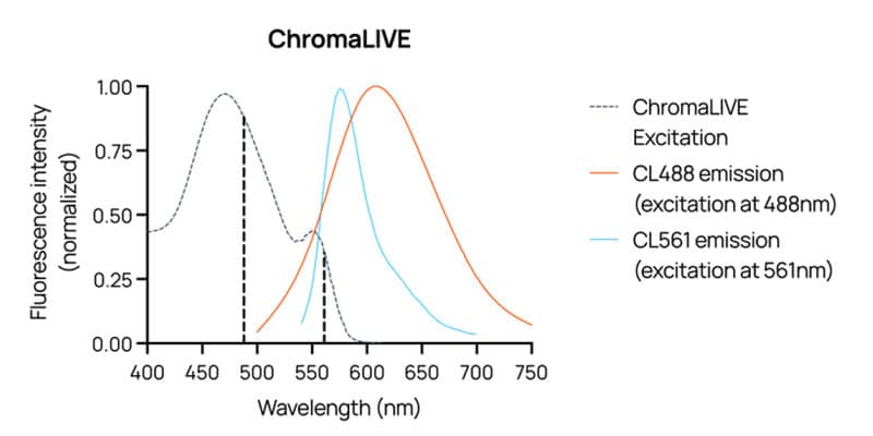
ChromaLIVE™ Non-Toxic Dye needs to be imaged at 2 different wavelengths minimally: ChromaLIVE561, and either ChromaLIVE488_Yellow or ChromaLIVE488_Red. That is because ChromaLIVE488_Yellow and ChromaLIVE488_Red look mostly similar (see figure 3), but can still provide slightly different information. However, selecting only one of these two is sufficient for differentiating between cellular phenotypes, even when phenotypes only have subtle differences
- 3 wavelenghts (Optional)
ChromaLIVE™ Non-Toxic Dye can be imaged at 3 different wavelengths: If possible, both ChromaLIVE488_Yellow and ChromaLIVE488_Red can be used
ChromaLIVE™ Non-Toxic Dye wavelengths:
- ChromaLIVE488_Yellow: excitation at 488 nm, image acquisition between 550-630 nm
- ChromaLIVE488_Red: excitation at 488 nm, image acquisition between 630-750 nm
- ChromaLIVE561: excitation at 561 nm, image acquisition between 590-630 nm

4. Image Examples


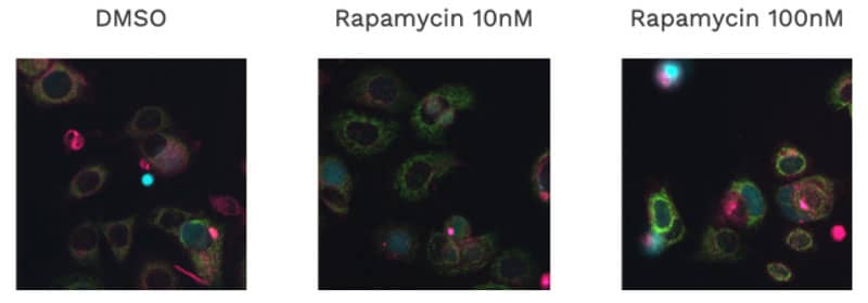
Figure 5. MCF-7 cells with ChromaLIVE™ Non-Toxic Dye at 72h treated with Rapamycin. Images are composite images of cells treated for an autophagy phenotype at 10 nM and 100 nM of rapamycin, in comparison with an untreated DMSO control. Red: ChromaLIVE 488-Yellow, Magenta: ChromaLIVE 488-Red, Green: ChromaLIVE 561-Yellow, Blue:Hoechst 33342.
5. Standard Protocol (for kinetic, 2D, live-cell assay)
MCF-7 cells, with standard compounds for apoptosis, ER stress and autophagy.
ChromaLIVE™ Non-Toxic Dye dilution and preparation (day0):
- Warm up the ChromaLIVE™ Non-Toxic Dye tube to room temperature before use to avoid condensation to form and water to get into the anhydrous dye solution
- Gently spin the tube before use to collect any dye solution that may remain near the cap
- Dilute 10 μL ChromaLIVE™ Non-Toxic Dye in 10 mL culture medium (1:1000 the provided solution)
- Vortex thoroughly
NOTE: Culture medium here is RPMI 1640 complemented with 10% FBS and 1% Penicillin/Streptomycin
Cell culture protocol with ChromaLIVE™ Non-Toxic Dye (day0):
- Harvest and count MCF-7 cells
- Resuspend cells in prepared culture medium with ChromaLIVE™ Non-Toxic Dye at 80,000 cells/mL
- Seed 96 well plate with 100 μL cell suspension per well, for a final amount of 8,000 cells per well
- Incubate overnight at 37°C, 5% CO2.
(OPTIONAL) Hoechst labeling of nucleus, for cell segmentation (day1)
- Dilute Hoechst 33342 solution at 1 μg/mL in culture medium, add 12.5 μL per well for a final concentration of 100 ng/mL
- Incubate for at least 3h, at 37°C, 5% CO2, before imaging.
Compound preparation and testing (day1)
- Prepare dose response curves with 10x concentrations, maintaining constant vehicle solvent concentration
- Prepare negative controls with vehicle solvent (here, 0.1% DMSO)
- Distribute 12.5 μL of test compounds or controls per well
Imaging and data acquisition (day1-3)
Image 96 well plate at 3h, 6h, 24h and 48h after addition of test compounds
Materials:
- 96-well plate: Greiner Bio-One Black μClear 96-well cell-culture treated plate, ref: 655090
- Nuclear stain: Hoechst 33342 (Invitrogen, ref: H1399)
- Vehicle: DMSO (Fisher BioReagents, ref: BP231-100)
- Culture medium: RPMI 1640 (ATCC modified) (Gibco, ref: A1049101), complemented with 10% FBS and 1% Penicillin/Streptomycin
6. Additional Information
Examples of acquisition channels and of filter settings:
- ChromaLIVE488_Yellow
- Excitation: 488 nm laser or 475/34 nm excitation filter
- Acquisition: 593/40 nm emission filter
- ChromaLIVE488_Red
- Excitation: 488 nm laser or 475/34 nm excitation filter
- Acquisition: 692/40 nm emission filter
- ChromaLIVE561
- Excitation: 561 nm laser or 560/32 nm excitation filter
- Acquisition: 593/40 nm emission filter
- Optional: DAPI
- Excitation: 405 nm laser or 377/54 nm excitation filter
- Acquisition: 447/60 nm emission filter
7. Control Compound Examples
| Cell death mechanism |
Control compounds (Concentration range, 1:10 serial dilution) |
End-point | Time points |
|---|---|---|---|
|
Apoptosis
|
Actinomycin D (1 pM-1 μM) Staurosporine (5 pM-5 μM) |
Actinomycin D: 72h Staurosporine: 24h |
12h, 24h, 48h, 72h 3h, 6h, 12h, 24h |
| ER stress |
Tunicamycin (10 pM-10 μM) Thapsigargin (1 pM-1 μM) |
Tunicamycin: 24h Thapsigargin: 24h |
3h, 6h, 12h, 24h 3h, 6h, 12h, 24h |
| Autophagy | Rapamycin (10 pM-10 μM) | Rapamycin: 72h | 12h, 24h, 48h, 72h |
Table 2. Doses and duration for MCF-7 cells in 2D
*Only provided as examples. Controls require validation.
**Images could be collected more frequently with the appropriate equipment, especially for time-lapse imaging (controlled temperature and CO2, auto-focusing, etc.)
ChromaLIVE™ is a registered trademark of Saguaro Biosciences.