Quantifying LNP Encapsulation Efficiency with RiboDyEE™
This is intended as a guide only.
In Brief Download the PDF of this protocol
This protocol describes the steps to measure RNA encapsulation inside a lipid nanoparticle (LNP) and quantify the total RNA concentration of samples using an ultra-sensitive RNA dye (RiboDyEE™ Cat. No. 9000).
1. Materials and Equipment
- RiboDyEE™ RNA Reagent (#9000)
- RNA standard (same as encapsulated in LNP)
- Triton X-100 (molecular biology grade)
- 20X TE buffer (molecular biology grade)
- Ultra-pure, nuclease free water
- Black, 96-well assay plate with lid (non-treated)
- Formulated LNP samples
- Fluorescence microplate reader
- Incubator
- Multichannel and single channel pipettes (2 – 200 μL)
- Filter pipette tips (2 – 200 μL)
- Petri dishes, or basins
- RNaseZAP™ RNase decontamination solution
2. Notes Before Starting
2.1 We provide RiboDyEE™ pre-aliquotted to minimise handling and the amount of freeze thaw cycles of the product. Keep aliquots stored at -20°C/-4°F when not in use and allow to thaw gently before performing the assay.
2.2 37 µL of RiboDyEE™ allow for the screening of 7 samples (+ 1 blank and calibration curves), in triplicate in the format described below. RiboDyEE™ 120 µL aliquots enable the encapsulation efficiency assay of a total of 21 samples, in triplicate, across 3 different plates.
2.3 To prevent RNase contamination of the RiboDyEE™ reagent and kit components, as well as all components used to prepare buffers:
- Clean working area by spraying RNaseZAP™ RNAse decontamination solution directly onto surfaces and wiping. Wait 5 minutes before re-cleaning with 70% ethanol solution.Spray
- RNaseZAP™ RNase decontamination solution onto a paper towel and surface clean pipettes and other lab apparatus that will be used throughout the procedure.
- If used, clean glassware with RNaseZAP™ RNase decontamination solution, allowing it to touch all surfaces by swirling or vortexing. Discard the solution and rinse twice with ultra-pure, nuclease-free water.
- Use clean, disposable gloves while handling all materials and avoid touching potentially contaminated surfaces.
- Use sterile, RNase free, filter pipette tips and plasticware.
3. Procedure
3.1 Preparation of buffers
3.1.1 TE buffer preparation: TE buffer (10 mM Tris-HCl, 1 mM EDTA, pH 7.5) is used for diluting the RiboDyEE™ reagent, LNP samples and ribosomal RNA standard solution. Dilute 20X TE buffer to 1X using ultra-pure nuclease-free water and mix by inversion.
3.1.2 Triton-TE buffer preparation: Prepare a 2% Triton-TE buffer using Triton X-100 and 1X TE buffer.
Tip: Use a syringe and blunt needle to measure and dispense Triton X-100 accurately without dripping. Triton X-100 is a strong surfactant and will cause bubbles when agitated. Warm the mixture to aid solubilization of Triton X-100 and mix gently to reduce bubbling.
3.2 Standard curve preparation (+/- Triton)
3.2.1 Prepare the RNA standards: Calculate the dilution necessary with TE buffer to prepare RNA stock solutions at 4 µg/mL and 0.2 µg/mL from nucleic acid standard solution. Note: It is important to use the same RNA as encapsulated in LNPs to ensure accuracy of results.
3.2.2 Standard curve dilutions: Prepare a series of RNA concentrations in wells down Columns 7 to 12. Final concentration ranges, after a 1:1 dilution with Triton-TE or TE buffer, and a further 1:1 dilution with RiboDyEE™ working solution (section 3.5), will result in a calibration curve from 50 – 1000 ng/mL RNA for wells prepared with Triton-TE, and a calibration curve from 1.25 – 50 ng/mL RNA for wells prepared with TE buffer, as shown in Figure 1.
- To wells in columns 7,8 and 9, prepare RNA concentrations from the 4 µg/mL RNA stock solution according to Table 1 and Figure 1.
- To wells in columns 10, 11, and 12, prepare RNA concentrations from the 0.2 µg/mL RNA stock solution according to Table 2 and Figure 1.
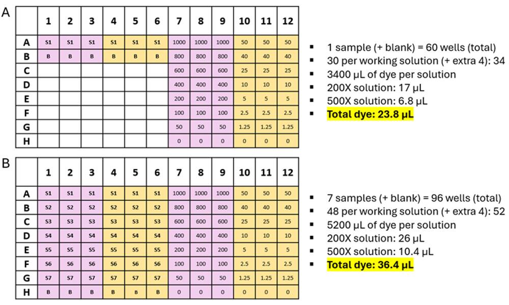
Figure 1. Experimental design of a RiboDyEE™ assay in a 96-well plate, pink wells are prepared with Triton-TE buffer, and yellow wells are prepared with TE buffer. Standard curves are plated in Columns 7 – 12 and the rest of the plate is used as needed for LNP samples. A) 1 sample (+ blank) is screened in triplicate. B) 7 samples (+ blank) are screened in triplicate.
Table 1. Volumes of RNA stock solution (4 µg/mL), TE buffer and Triton-TE required to plate final RNA concentrations as shown, accounting for a 1:1 dilution with RiboDyEE™ reagent.
|
Final RNA concentration (ng/mL) |
RNA stock solution (4 µg/mL) (µL) |
TE buffer (µL) |
Triton- TE (µL) |
|---|---|---|---|
| 1000 | 50 | 0 | 50 |
|
800 |
40 | 10 | 50 |
|
600 |
30 | 20 | 50 |
|
400 |
20 | 30 | 50 |
|
200 |
10 | 40 | 50 |
|
100 |
5 | 45 | 50 |
|
50 |
2.5 | 47.5 | 50 |
|
0 |
0 | 50 | 50 |
Table 2: Concentrations as shown, accounting for a 1:1 dilution with RiboDyEE reagent.
|
Final RNA concentration (ng/mL)
|
RNA stock solution (0.2 µg/mL) (µL) |
TE buffer (µL)
|
|---|---|---|
| 50 | 50 | 50 |
|
40 |
40 | 60 |
| 25 | 25 | 75 |
| 10 | 10 | 90 |
| 5 | 5 | 95 |
| 2.5 | 2.5 | 97.5 |
| 1.25 | 1.25 | 98.75 |
| 0 | 0 | 100 |
3.3 Preparing and plating LNP samples
3.3.1 Remove LNP samples from storage and allow to warm to room temperature before pipetting.
3.3.2 Prepare 350 μL of LNP samples at a concentration of 3 μg/mL RNA using TE buffer as a diluent. Calculate the volume of each LNP sample required for dilution using the theoretical RNA concentration. Note: The total concentration of LNP sample in sample wells should be within the standard curve range of 50 – 1000 ng/mL (see section 3.2). A target LNP concentration of 3 μg/mL accounts for the 1:1 dilution with TE or Triton-TE buffer (sections 3.3.5 and 3.3.6), and a further 1:1 dilution with RiboDyEE™ working solution (section 3.5) and will result in a final RNA concentration 750 ng/mL in the sample wells.
3.3.3 Prepare Blank samples (labelled B – see Figure 1) by diluting a volume of PBS/Tris, equivalent to that used in LNP sample preparation, to 350 μL using TE buffer.
3.3.4 Add 50 µL of each LNP sample to appropriate wells as shown in Figure 1. Each LNP sample (+/- Triton) is plated over three rows to give triplcate results.
3.3.5 Add 50 μL of Triton-TE buffer to the required number of wells in columns 1, 2 and 3. Gently mix the sample with buffer by pipetting. Tip: Do not remove the tip from the solution when mixing to prevent bubble formation.
3.3.6 Add 50 μL of TE buffer to the required number of wells in columns 4, 5 and 6. Gently mix the sample with buffer by pipetting.
3.4 Lyse the LNPs
Place the lid on the 96-well plate and incubate the 96-well plate for 15 minutes at 37 °C. Let the plate cool to room temperature before removing the lid.
3.5 Preparation and addition of the RiboDyEE working solutions.
3.5.1 Remove the RiboDyEE™ reagent from the fridge and allow to reach room temperature before use. Keep the vial protected from light. Note: The reagent must be a liquid consistency before opening the vial to prevent moisture absorption and resulting loss of reagent efficacy.
3.5.2 The RiboDyEE™ working solution is prepared by dilution of the reagent with TE buffer. To wells prepared using Triton-TE buffer, a 200X dilution of the RiboDyEE™ reagent is required, and for wells prepared using TE buffer, a 500X dilution is required.
- Count the number of wells in use. In Figure 1A, 60 wells are in use (30 w/Triton-TE and 30 w/TE buffer). Assume 4 extra wells per working solution.
- To ensure a 1:1 dilution with the well contents, 100 µL of RiboDyEE™ working solution is added to each well, and so 3400 µL of each working solution should be prepared. Vortex for 10 seconds to mix.
200X dilution: Add 17 µL RiboDyEE reagent to 3383 µL TE buffer.
500X dilution: Add 6.8 µL RiboDyEE reagent to 3393.2 µL TE buffer.
- Note: The RiboDyEE™ working solution should be prepared in sterile, disposable polypropylene plasticware rather than glassware as the reagent may adsorb to glass surfaces. It should also be protected from light and used within a few hours of preparation.
3.5.3 To wells prepared with Triton-TE buffer, add 100 µL of the 200X working solution, and to wells prepared with TE buffer, add 100 µL of the 500X working solution. Mix gently by pipetting.
3.5.4 If present, remove bubbles from wells using a blunt needle as these can affect fluorescent measurements.
3.6 Plate Analysis
3.6.1 Remove the lid and place the plate into the plate reader. Measure the fluorescence using a fluorescence microplate reader and the following parameters:
- Wavelength: excitation 485 nm, emission 525 nm
- Gain: 1400 (instrument dependent)
- Optic: Top
3.6.2 Prepare calibration curves from the RNA standards and determine the linear range and equation of the curve, LOD and LOQ.
3.6.3 Remove the appropriate ‘blank’ fluorescent values from each sample and use the calibration curves created with Triton-TE and TE buffers to convert sample fluorescence into ‘total’ and ‘free’ RNA concentrations, respectively. Incorporate sample dilutions to determine the ‘total’ and ‘free’ concentrations of the LNP samples.
3.7 Calculate the Encapsulation Efficiency (%EE) and Mass Balance %
3.7.1 Calculate the %EE using the equation below:
%EE = [(CT-CF/CT)] * 100
Where: CT = Total RNA concentration (based on results from the wells prepared with Triton-TE buffer)
CF = Free, unentrapped RNA concentration (based on results from the wells prepared with TE buffer)
3.7.2 Calculate the mass balance (also can be referred to as Recovery/yield) as follows:
Mass balance = [CT/Theoretical concentration of RNA] * 100
Where: CT = Total RNA concentration (based on results from the wells prepared with Triton-TE buffer
NB. Mass Balance should be between 90 and 110%.
Acknowledgements
This protocol has been developed and optimized by the lab of Professor Yvonne Perrie at Strathclyde Institute of Pharmacy and Biomedical Sciences and is used with their permission.